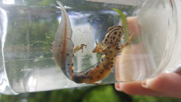
The Chair of Zoology and Microbiology is an organizational unit for research, knowledge transfer and professional issues in the field of animal biology, biodiversity and microbiology.
The mission of the chair is:
- development of new knowledge in the feeld of animal biology from the perspective of body organization comparismen , physiology and ethology;
- understanding of animal biodiversity and biogeography thrue studies of patterns and processes of animal adaptations to changing environmental conditions ;
- knowledge transfer through the development and implementation of our study programs;
- providing lifelong learning and professional upgrade in fields of animal biology for biology and science teachers;
- participation in working groups to solve professional issues in the field of animal biology and nature conservation as well as issues within private, public, and governmental institutions;
- participation in professional association and in public discussions in the field of animal biology, biodiversity, and conservation;
at the fild of Microbiology, we study microorganisms from various natural and industrial environments. We use classical microbiological tools, modern molecular-biological methods, and bioinformatics to explore their taxonomic and metabolic diversity. From selected strains of bacterial species, we isolate and characterize industrially interesting products.
Morphology, physiology and genomics of bacteria from different environmental niches
In addition to viruses, bacteria are the most widespread group of organisms on Earth. Due to their small size and short generation time, they can quickly adapt to new environmental conditions by diversifying metabolic pathways and cell forms. Identification of new species, analysis of their genomes and adaptation mechanisms are the contents of this research area.
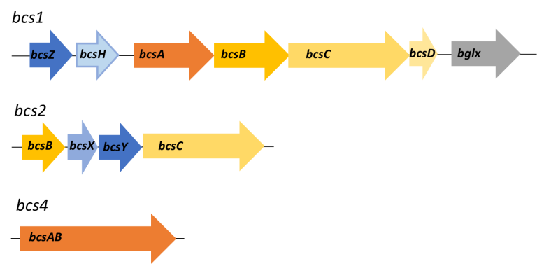
Figure 1: The bacterium Komagataeibacter melomenusus has different operons for nanocellulose synthesis (Marič et al., 2020).
Extracellular polysaccharides: from genes to product
Microorganisms secrete various polysaccharides into their exterior to protect the cell from drying out, from the intrusion of inhibitory substances, allowing the cells to attach to various surfaces or to float on the surface of liquids. These extracellular polysaccharides are also useful in medicine (preparation of replacement tissues, capillaries, patches) and in the food industry (emulsifiers and food stabilizers). Our research is focused on the isolation and characterization of acetan, acetan-like polysaccharides and nanocellulose produced by acetic acid bacteria, and their application for various medical and food products.
Gorgieva S. and Trček J. 2019. Bacterial Cellulose: Production, Modification and Perspectives in Biomedical Applications. Nanomaterials 9(10):1352. doi: 10.3390/nano9101352.
Škraban J., Cleenwerck I., Vandamme P., Fanedl L., Trček J. 2018. Genome Sequences and Description of Novel Exopolysaccharides Producing Species Komagataeibacter pomaceti sp. nov. and Reclassification of Komagataeibacter kombuchae (Dutta and Gachhui 2007) Yamada et al., 2013 as
a Later Heterotypic Synonym of Komagataeibacter hansenii (Gosselé et al. 1983) Yamada et al., 2013. Syst Appl Microbiol 41(6):581-592; doi: 10.1016/j.syapm.2018.08.006.
Škraban J. and Trček J. 2017. Comparative Genomics of Acetobacter and Other Acetic Acid Bacteria. In: Acetic Acid Bacteria: Fundamentals and Food Applications, p. 44-70. Editor: Ilikin Yucel Sengun, CRC Press.
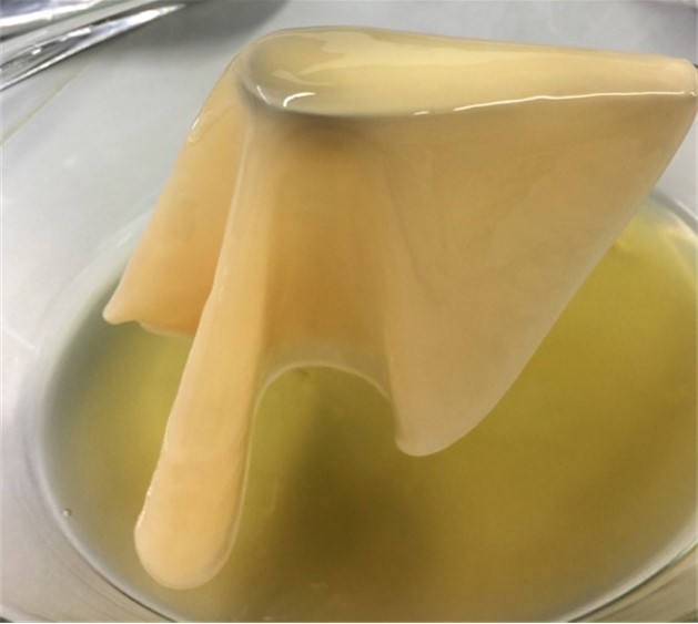
Figure 2: Bacterial species Komagataeibacter melomenusus is an efficient producer of nanocellulose (Marič et al. 2020).
Investigating cell function under normal and pathophysiological conditions
The group is working on several lines of research:
- Mechanism of action of pancreatic endocrine and exocrine cells: beta cells, alpha cells, ductal cells, acinar cells.
- Mechanism of action of adrenal chromaffin cells:
- Testing pharmacological substances that are candidates for new drugs.
- Alterations and causality of endocrine cell changes in animal models of diet-induced diabetes
- Alterations and causality of ductal cell changes and their relationship to pancreatic endocrine cells, under conditions of pancreatitis
Techniques used to investigate cell function:
Confocal recording of intracellular calcium ([Ca2+]i) concentration dynamics.
Long time series allow the observation of long-lasting [Ca2+]i dynamics, and high-frequency (-50 Hz) image acquisition provides good temporal resolution of [Ca2+]i dynamics in beta cells.
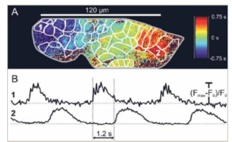
Figure 1: Confocal measurement of intracellular calcium concentration dynamics in beta cells. A: Calcium dynamics in the islets of Langerhans are organised in the form of waves propagating across the islet. The colours indicate the time lag of the oscillations of the intracellular calcium concentration. B: Temporal dynamics of intracellular calcium concentration in two beta cells after stimulation with 12 mM glucose.y
Patch clamp method:
this method determines the electrical activity in the beta-cells, and furthermore the ion channel activity can be evaluated. The method closely supports the Ca measurements.
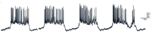
Figure 2: Membrane potential dynamics of the beta cell during stimulation with 12 mM glucose. Cells are rhythmically activated, superimposed rapid changes in potential are visible.


